Eyelid Bump Removal And Testing
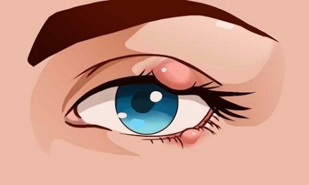
 Eyelid bumps are painful red lumps that appear at the edge of the eyelid. This occurs where the lash meets the eyelid. Blockage or bacteria in the oil glands of the lid is the reason for most bumps. Eyelid bumps don’t usually require medical attention since they are harmless.
In most cases, bumps go away with basic attention. However, you should speak to your doctor if it begins interfering with your vision or stops responding to home treatments. Your doctor will look for signs of other serious problems and ways to manage the symptoms.
Khan Eyelid and Facial Aesthetics, led by board certified ophthalmologist Dr. Tanya Khan, provides safe and proven eye care procedures to patients in Plano, Dallas, Texas, and surrounding locations.
Different Types of Eyelid Bumps
Eyelid bumps are of three common types. The underlying cause and type of your eyelid bump is going to determine the best treatment course.
Styes
Styes are the most common eyelid bump type. These occur when bacteria get into the eyelid oil glands. Styes are described as a round, red bump which may occur close to the eyelash. They can cause your eyelid to feel sore.
Chalazion
Chalazion is an inflammatory lesion and grows further than a stye does. They usually occur due to a blockage in the tear or oil-producing glands of the eyelids. Chalazion may interfere with your vision depending on how big it gets or where it grows.
Xanthelasma
Xanthelasma are usually an indicator of high cholesterol levels and occur mostly in older adults. They are characterized as harmless, yellow bumps, which occur with fat buildup under the eye skin.
Papillomas
A pedunculated papilloma on the upper eyelid.Papillomas are the most common benign lesion of the eyelid. These growths represent a benign hyperplasia of the surface epithelium and may be sessile (flat) or pedunculated (on a stalk). They occur in middle aged and elderly individuals and may be solitary or multiple, occurring anywhere on the eyelid. Papillomas differ from the infective warts which consist of inflammatory hypertrophy of the surface epithelium. Treatment is by surgical excision.
Syringomas
Syringomas on the left lower eyelid.Syringomas are benign, skin coloured papules 2-3 mm in diameter. They are usually located on the lower eyelids and cheeks but can be found on the upper lids. They mainly occur in women at any age after the mid teens. They involve the full thickness of the skin and so will recur if not completely excised.
Seborrhoeic Keratosis
An area of seborrhoeic keratosis in the middle of the right lower eyelid.Seborrhoeic keratosis is a common benign lesion on the lids of aging individuals.
They are well circumscribed, waxy, friable and appear stuck on to the skin. The lesion is very superficial and may be pigmented. Microscopically, they show hyperkeratosis and cystic areas filled with keratin. Treatment involves surgical excision.
Naevi
A naevus in the inner corner of the left lower eyelid.Naevi are common in the eyelid area and may be pigmented or non-pigmented. The
naevus consists of a collection of benign appearing dermal melanocytes. Generally, they are present for years with no change. If the duration of any pigmented
lesion is unknown, acquired with age (especially after 40) or there is a change in size or colour, a biopsy is recommended.
Hordeola (Styes) and Chalazions
An internal hordeolumAn external hordeolum (stye) results from an acute purulent inflammation of the superficial sweat glands, sebaceous glands or hair follicle of the eyelids, while an internal hordeolum occurs in the meibomian glands within the tarsal plates of the lids.
A chalazion is a chronic inflammation of a meibomian gland (deep type) or Zeiss sebaceous gland (superficial A chalaziontype) resulting in a clinically firm, painless nodule of the eyelid.
Treatment of the acute phase may involve hot compresses, antibiotics (drops, ointment, or tablets) while treatment of the chronic phase usually involves incision and drainage of the cyst.
Epidermal Inclusion Cysts
Epidermal inclusion cysts.These are small white-yellow cystic lesions occurring on the lid skin, conjunctiva, face or neck and are very common. They may develop spontaneously or arise following trauma or after surgery along an incision line. They originate from pilosebaceous follicles or implantation of surface epidermis. Some of these cysts occurring amongst the lashes will be difficult to distinguish from a blocked Zeiss gland (sebaceous gland). Treatment involves excision, and if the cyst wall is not removed, recurrence may occur.
Sebaceous Cysts
Sebaceous cyst of the left lower eyelid.Sebaceous cysts occur around the eyelid area and clinically resemble epidermal inclusion cysts. They are generally found in locations with many hair follicles, particularly the brow area and medial canthus. These cysts may occur secondary to obstruction of the Zeiss gland, meibomian gland or sebaceous glands associated with hair follicles of the lid skin or brow area. Unlike an epidermal inclusion cyst (filled with keratin material), a sebaceous cyst contains epithelial cells, keratin, fats and cholesterol crystals. Treatment involves surgical excision.
Milia
Milia are multiple well-delineated, round, yellow-white cystic lesions ranging from 1 to 3 mm in diameter, found on the face, lids, cheeks and nose. They may occur spontaneously or arise following trauma. They are felt to be retention follicular cysts caused by blockage of the fine pilosebaceous units (hair follicle). Surgical excision is the treatment of choice.
Retention Cysts, Cysts of Moll
Cysts of Moll on the right upper and lower eyelids.The lid skin has numerous sweat glands (eccrine glands) and modified sweat glands (apocrine glands) such as the glands of Moll. Blockage of these glands leads to the formation of a translucent (water blister-like) lesion on the lid skin or lid margin amongst the lashes. They may occur as a single lesion or as several. Treatment involves total excision otherwise they will recur. Simply stabbing them with a needle is ineffective in allowing their resolution.
Eyelid Bump Treatment Options
Eye doctors can diagnose chalazion or stye by looking at it. They may flip your eyelid over quickly to have a closer look depending on where the eye bump is located. Generally, medical tests are not necessary unless there is a concern that the medical problem is something different.
Basic home care
You should never try popping or squeezing a chalazion or style. This can increase the risk of bacteria spread and infection to the other eye. Instead, you can hold a warm compress to the stye for about 10 minutes. This can be done for up to four times a day. Compression and heat can go a long way in draining the stye and loosening any oil gland blockages. This helps in overall healing. You don’t need home care for Xanthelasma.
Medical care
Your doctor may need to puncture and drain the infected fluid in large styes. They may prescribe an antibiotic cream to be applied to your eyelid if you keep getting frequent styes or have a stubborn. Surgery is usually reserved as an option in the case of large chalazions that don’t go away.
You would be given antibiotic drops to be used before and after the surgery for treating and preventing the infection. The doctor may also administer anti-inflammatory steroid injections to relieve swelling. Oculoplatic & reconstructive surgeon Dr. Tanya Khan receives patients from Plano, Dallas, Texas, and nearby areas for advanced and effective eye care treatments.
Contact Khan Eyelid and Facial Aesthetics and Oculoplastic & Reconstructive Surgeon Dr. Tanya Khan Today to Schedule an Appointment
For more information about procedures and treatments at Khan Eyelid and Facial Aesthetics by Ophthalmic surgeon Dr. Tanya Khan. Click here to contact us.
Taking patients from in and around Dallas, Plano, Fort Worth, Grapevine, Garland, Mesquite, Carrollton, Irving, Frisco, Texas and more.


Schedule a Consultation: 972-EYE-LIDS (393-5437)